
Chest X-Ray Dataset
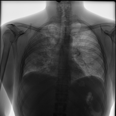
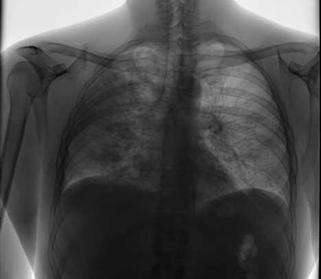
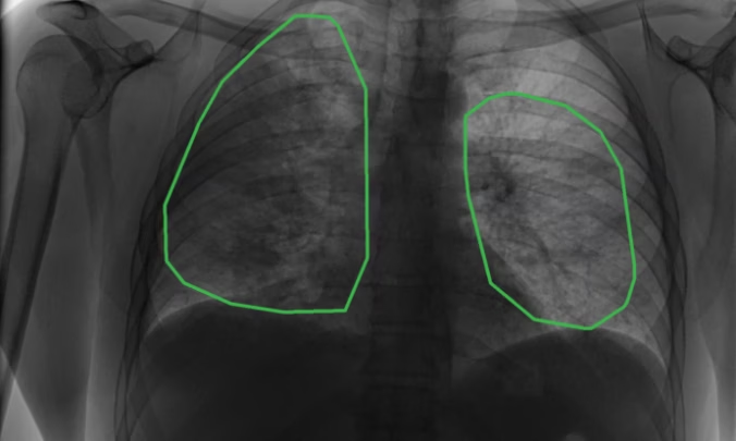
The NIH chest X-ray dataset contains labeled chest radiographs with detailed segmentation of pathologies, providing high-quality X-ray images of chest for detecting lung diseases, pulmonary conditions, and other medical imaging tasks, making it a valuable resource for diagnostic imaging, computer vision, and deep learning in healthcare
-
- Files
- 443
-
- Medical Studies
- 150
-
- Data tags
- 13
- Medicine
- Computer Vision
- Segmentation
- Classification
- Machine Learning
The NIH chest X-ray dataset contains labeled chest radiographs with detailed segmentation of pathologies, providing high-quality X-ray images of chest for detecting lung diseases, pulmonary conditions, and other medical imaging tasks, making it a valuable resource for diagnostic imaging, computer vision, and deep learning in healthcare
- Medicine
- Computer Vision
- Segmentation
- Classification
- Machine Learning
-
- Files
- 443
-
- Medical Studies
- 150
-
- Data tags
- 13
Dataset Info
| Characteristic | Data |
| Description | Chest X-ray to recognize pathologies |
| Data types | DiCOM |
| Markup | Segmentation of pathologies |
| Tasks | Pathology recognition, computer vision. |
| Total number of files | 443 |
| Number of studies | 150 |
| Labeling | ‘Nodule/mass’, ‘Dissemination’, ‘Annular shadows’, ‘Petrifications’, ‘Pleural effusion’, ‘Pneumothorax’, ‘Rib fractures’, ‘Healed rib fracture’, ‘Atelectasis’, ‘Enlarged mediastinum’, ‘Hilar enlargement’, ‘Infiltration/Consolidation’, ‘Fibrosis’ |
| Gender | Male, female |
| Age | 25 - 70 |
Statistics
-
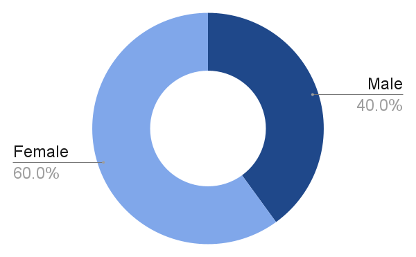
- Distribution by gender
-
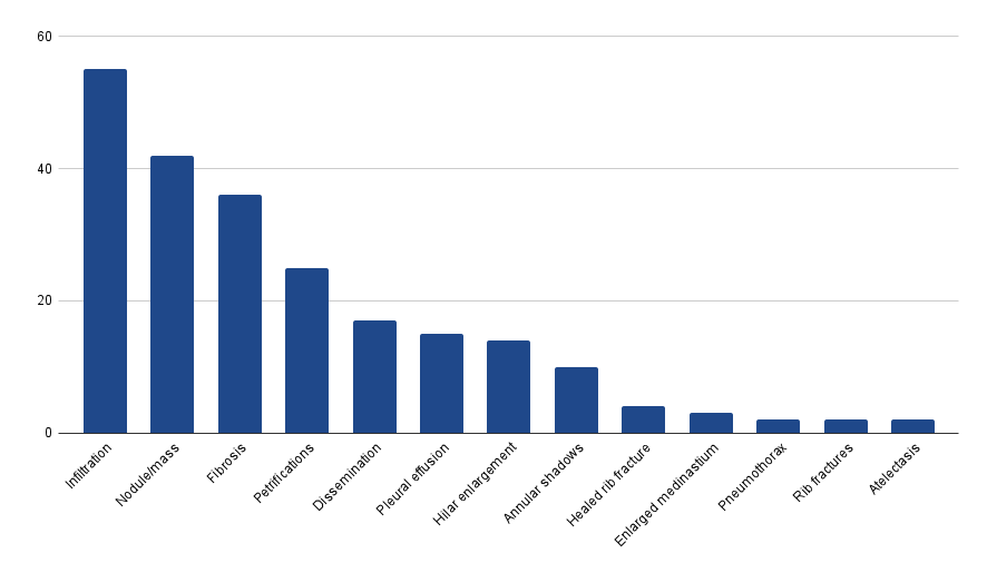
- Number of studies for each condition
Technical
Characteristics
| Characteristic | Data |
| File extension | DiCOM |
| Markup format | JSON |
Dataset Use Cases
FAQs
Unidata Cases

Digital Tree Passport Annotation for Forest Mapping
- Forestry Monitoring & GIS
- 2 months
- 200,000 trees, 10 species classes
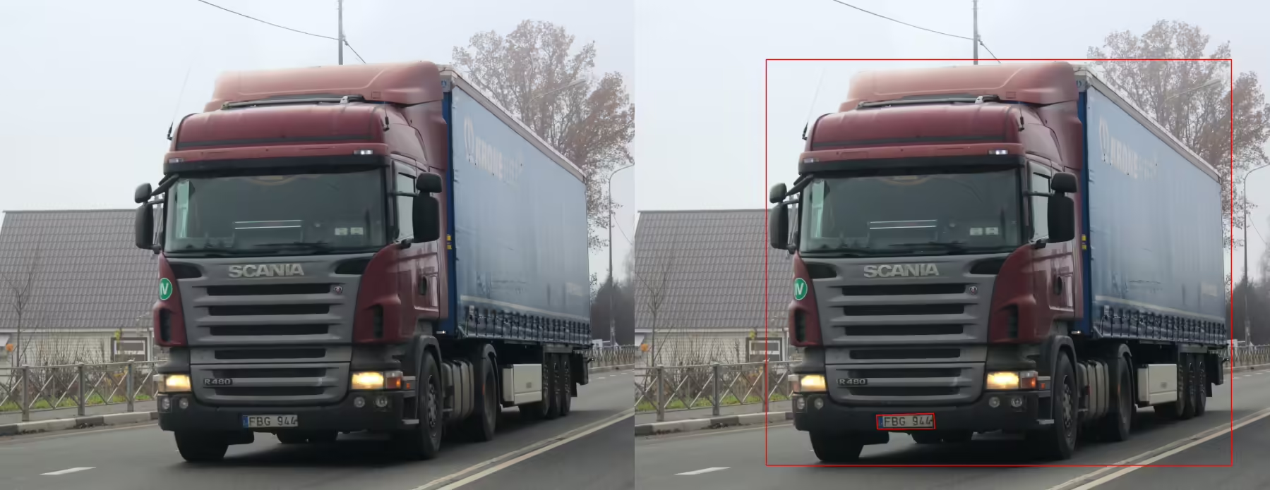
License Plate Annotation for Vehicle Recognition System
- 100,000 images with detailed license plate markup (bounding boxes, digits, regional symbols)
- 2 weeks
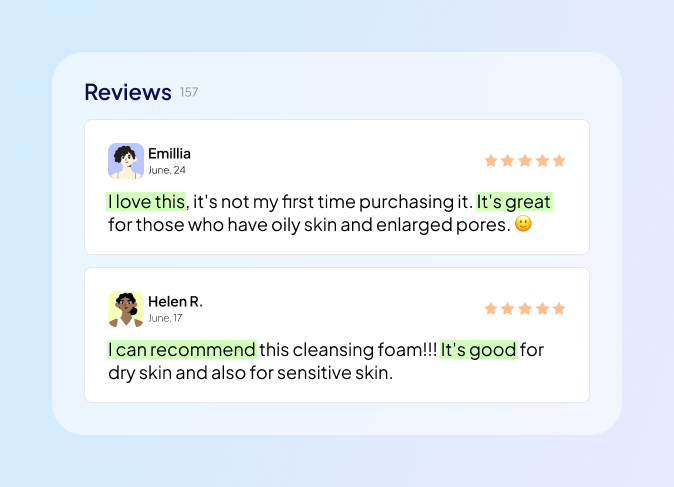
Sentiment Annotation for Brand Monitoring
- Marketing & Consumer Insights
- 12,000 text samples, 3 sentiment classes (positive, negative, neutral)
- 3 weeks

Surveillance Video Annotation for Entrance Monitoring
- Surveillance & Security
- 90 minutes of video from three cameras, approximately 50-60 thousand frames
- 2 week
Similar Datasets
-
 Commercial
- Medicine
- Classification
- Machine Learning
- Computer Vision
- Data Labeling
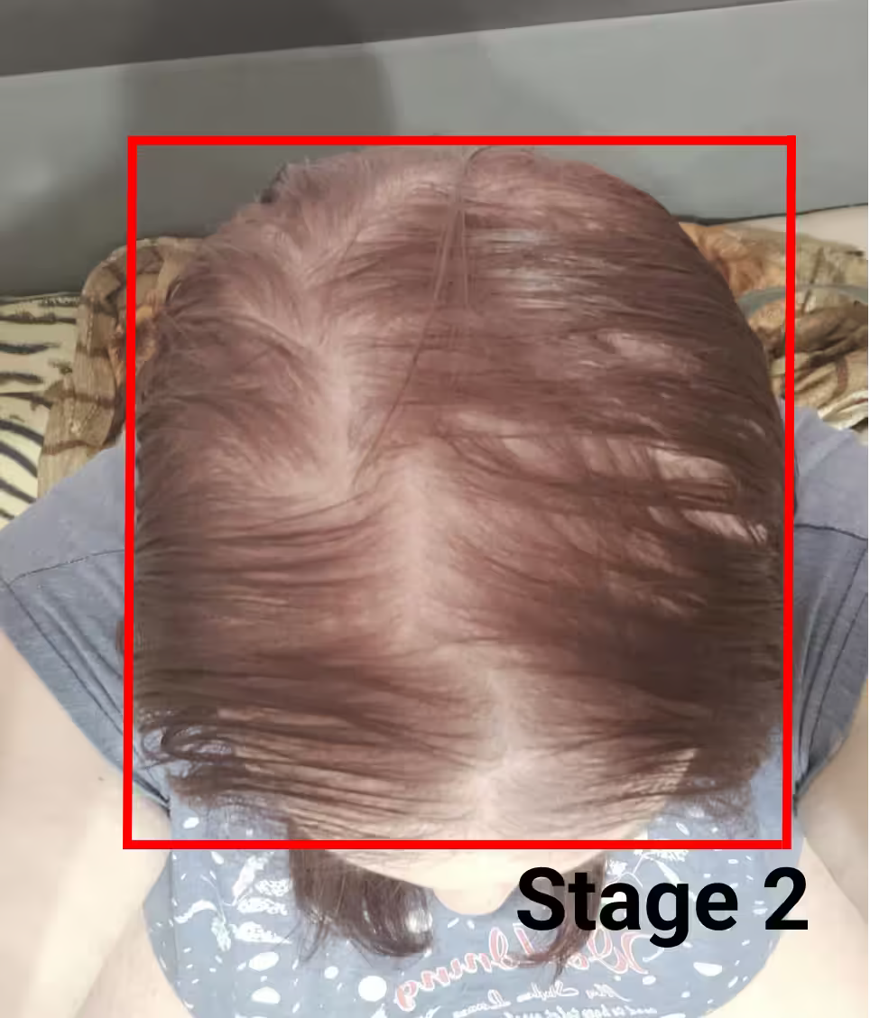
Female Hair Loss Dataset
This hair loss dataset provides alopecia images of women captured from the top and front sides, labeled into three classes based on the Ludwig scale, with metadata (age, gender, ethnicity) to support machine learning, deep learning, and AI models for hair loss detection, early diagnosis, and research on hair thinning and scalp health.
552 Images
276 People -

 Commercial
- Medicine
- Classification
- Computer Vision
- Machine Learning
- Segmentation
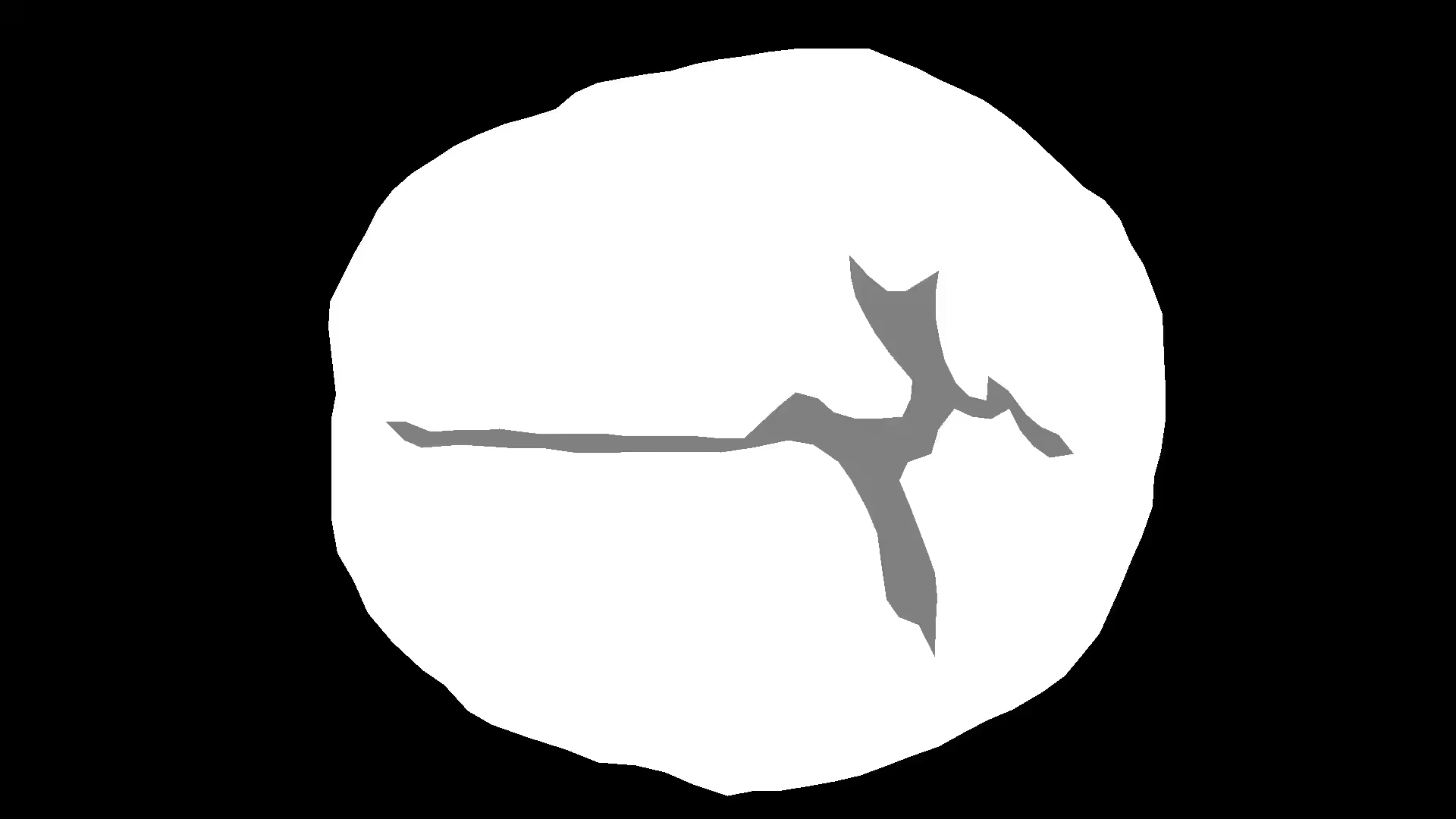
Men Hair Loss Segmentation Dataset
Men Hair Loss Segmentation Dataset is an accurately labeled bald men dataset consisting of multi-angle images of individuals with varying degrees of hair loss, labeled for alopecia classification, and designed to support machine learning models in analyzing scalp conditions, hair thinning, and developing hair restoration and hair transplantation solutions using advanced deep learning techniques.
3 100 Images
775 People -
 Commercial
- Computer Vision
- Medicine
- Classification
- Machine Learning
- Segmentation
Women Hair Loss Segmentation Dataset
This hair loss dataset features annotated images of women with varying degrees of hair loss, designed for machine learning and deep learning applications focused on analyzing hair density, scalp conditions, and developing solutions for hair restoration, hair thinning, and alopecia treatment
108 Images
54 People -
 Commercial
- Computer Vision
- Medicine
- Classification
- Machine Learning
- Data Labeling
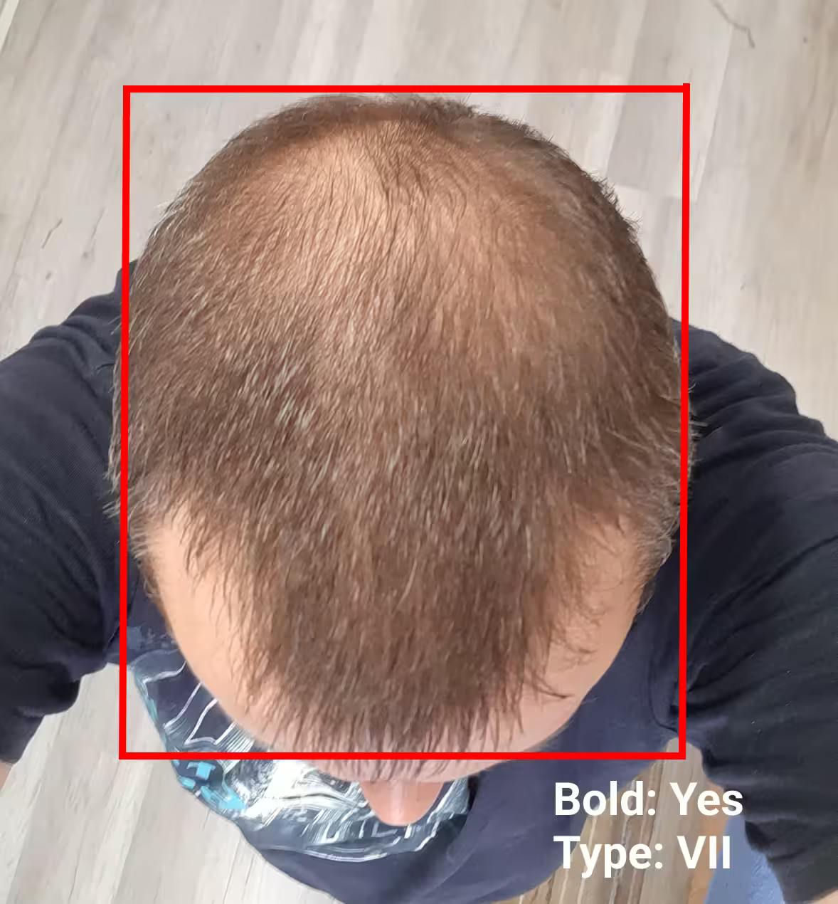
Male Hair Loss Dataset
The hair loss dataset contains high-resolution scalp images of people captured from five sides, labeled with seven classes on the Norwood-Hamilton scale and supplemented with hair follicle annotations to help machine learning models analyze hair density, thinning patterns, and diagnose baldness
2 260 Images
452 People -
 Commercial
- Medicine
- Computer vision
- Machine Learning
- Segmentation
- Classification
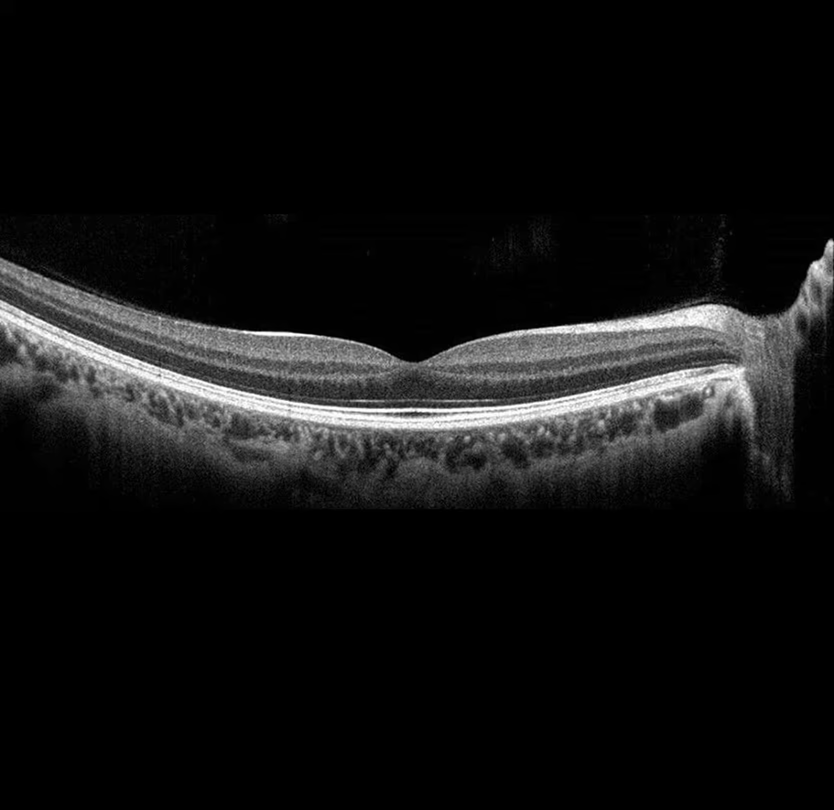
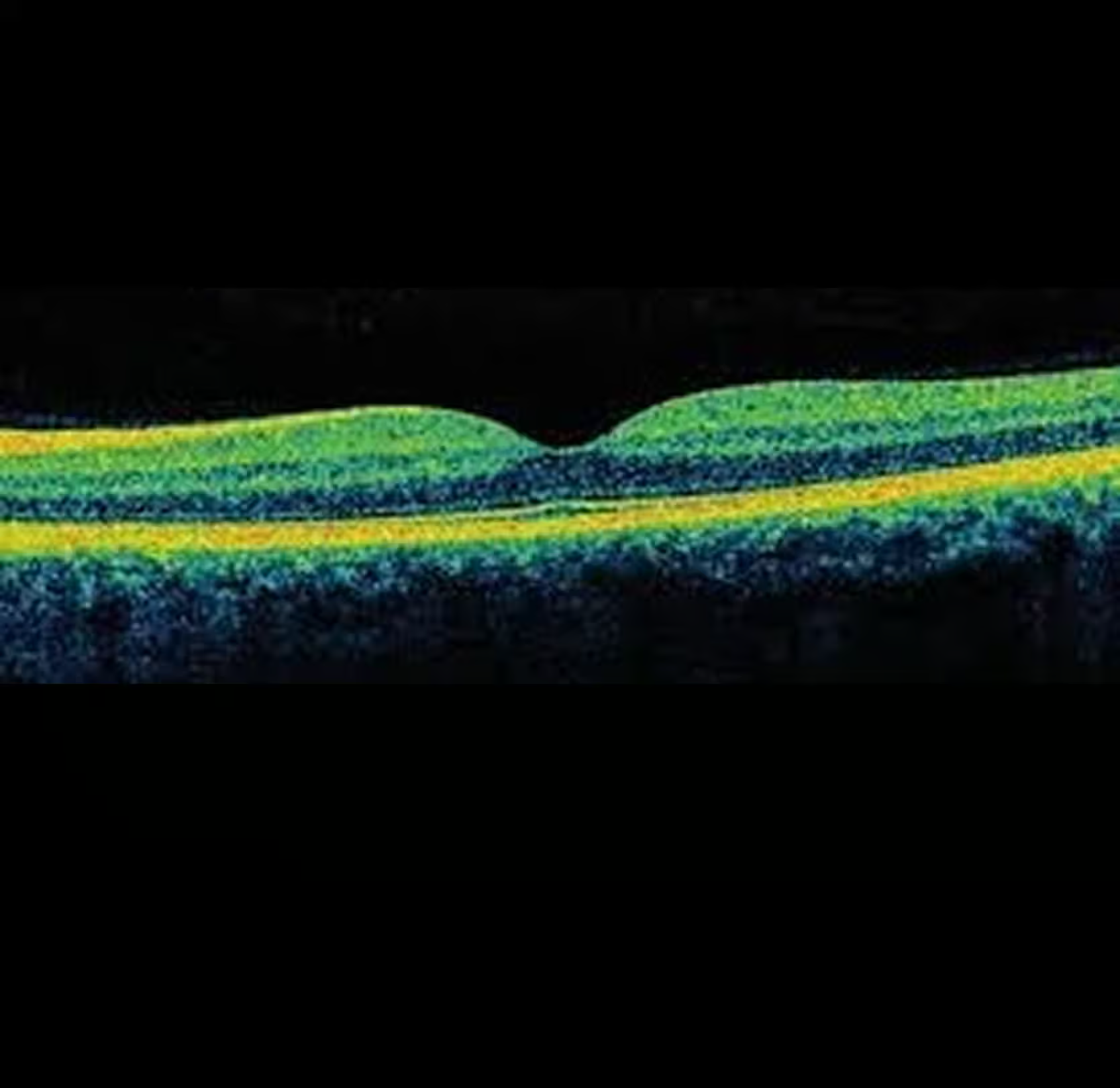
OCT Retinal Dataset
OCT Retinal Dataset features 1,000 volumetric OCT scans (B-scans) of the retina with annotated layers, fluid regions, and clinical diagnoses, including AMD, diabetic retinopathy, and glaucoma. This DiCOM dataset supports AI model training in disease classification, pathology segmentation, and computer vision–based retinal analysis using coherence tomography images.
1,000 Scans
100 B-scans per volume -
 Commercial
- Medicine
- Computer vision
- Machine Learning
- Segmentation
- Classification
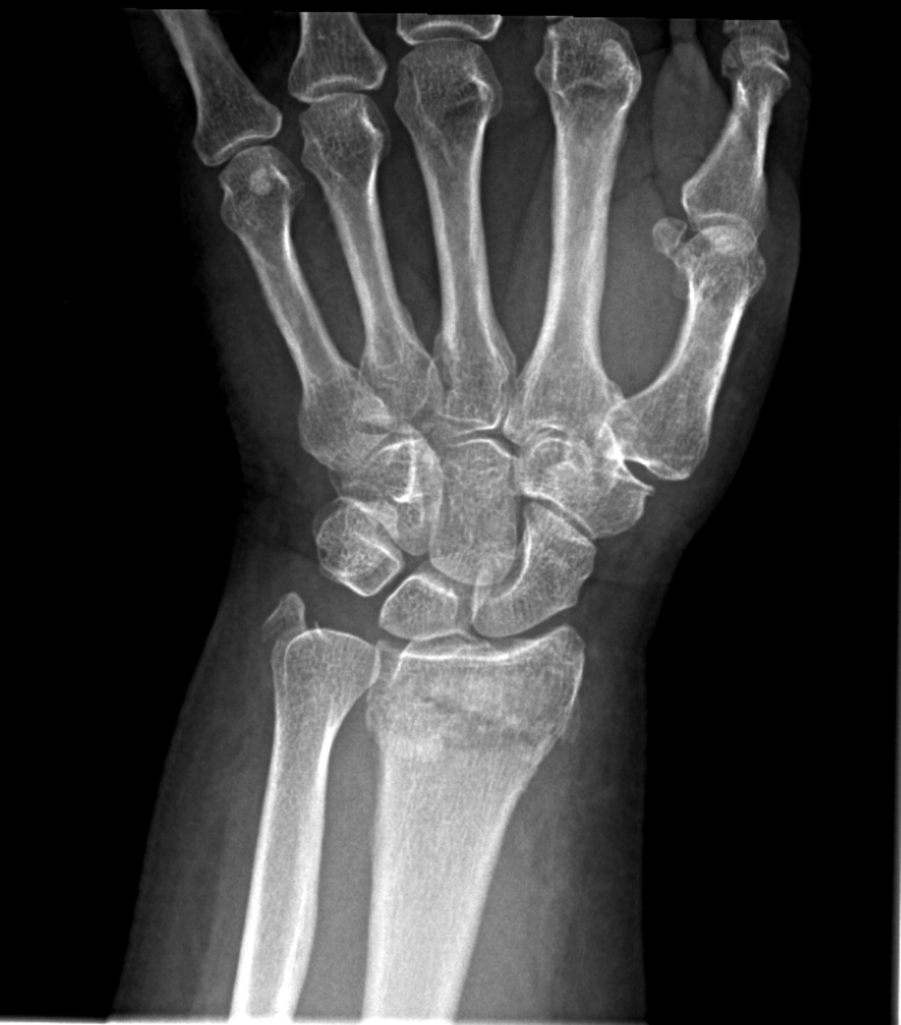
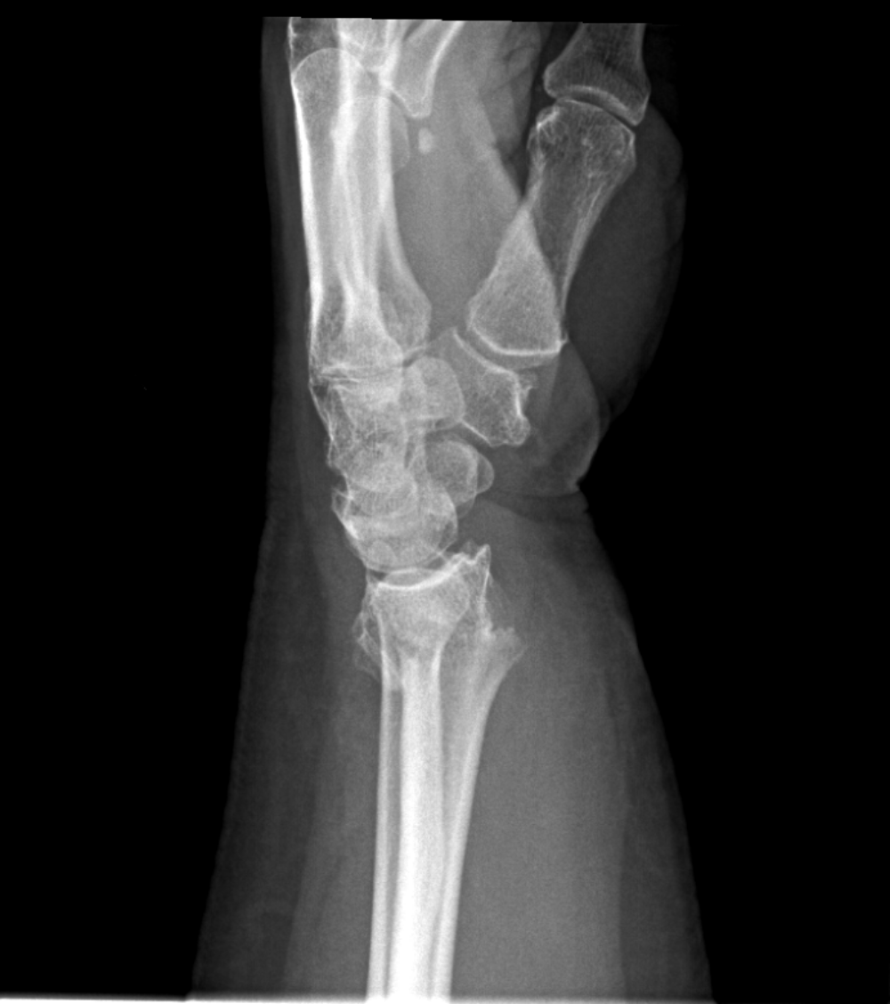
X-ray of upper-extremity joints
This large-scale X-ray labeled images dataset comprises over 1.2 million digital X-ray images focused on the shoulder, elbow, and wrist joints, designed for pathology recognition, segmentation tasks, and training deep learning models in medical imaging, with detailed annotations for the accurate detection of bone and joint conditions.
200,000+ studies with protocol
1,000,000+ studies without protocol
13 pathologies -

 Commercial
- Medicine
- Computer vision
- Machine Learning
- Segmentation
- Classification
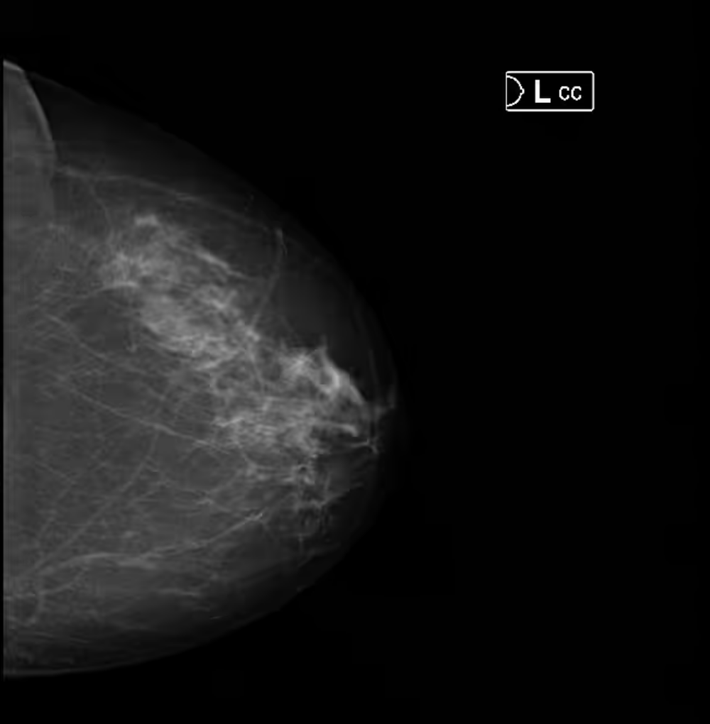
Mammography Dataset
Mammography Dataset is a comprehensive DDSM dataset containing over 600,000 digital mammograms with pathology segmentation and study-level labels, designed for cancer detection, breast imaging, and training AI models in cancer diagnosis using real-world mammographic images from screening mammography exams.
100,000+ studies with protocol
500,000+ studies without protocol -
 Commercial
- Medicine
- Computer vision
- Machine Learning
- Segmentation
- Classification
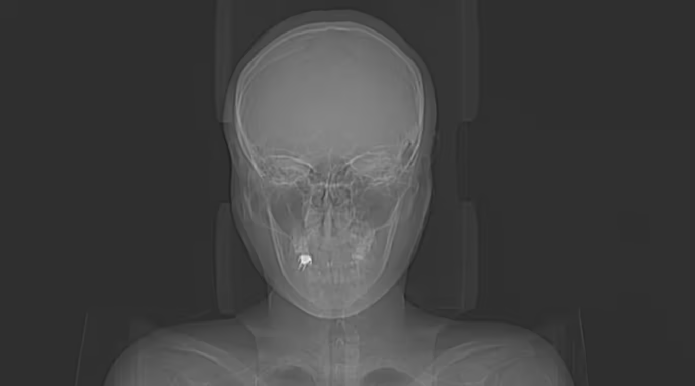
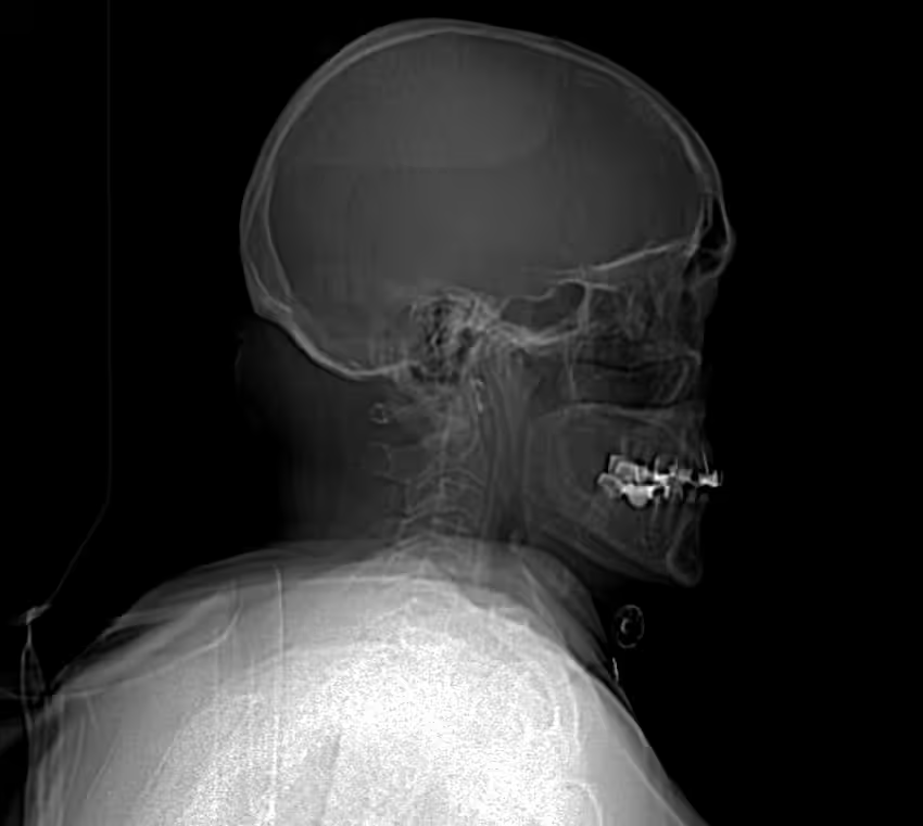
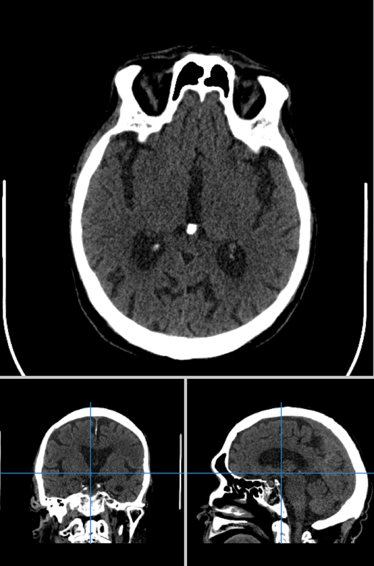
CT Scans of Brain
This brain CT dataset comprises over 70,000 DICOM studies with labeled pathologies such as intracerebral hemorrhage, ischemic stroke, and vessel abnormalities, providing segmentation data and annotations to support medical research, computer vision, and deep learning–based diagnostic algorithms.
20,000+ studies with protocol
50,000+ studies without protocol
5 pathologies -
 Commercial
- Medicine
- Computer vision
- Machine Learning
- Segmentation
- Classification
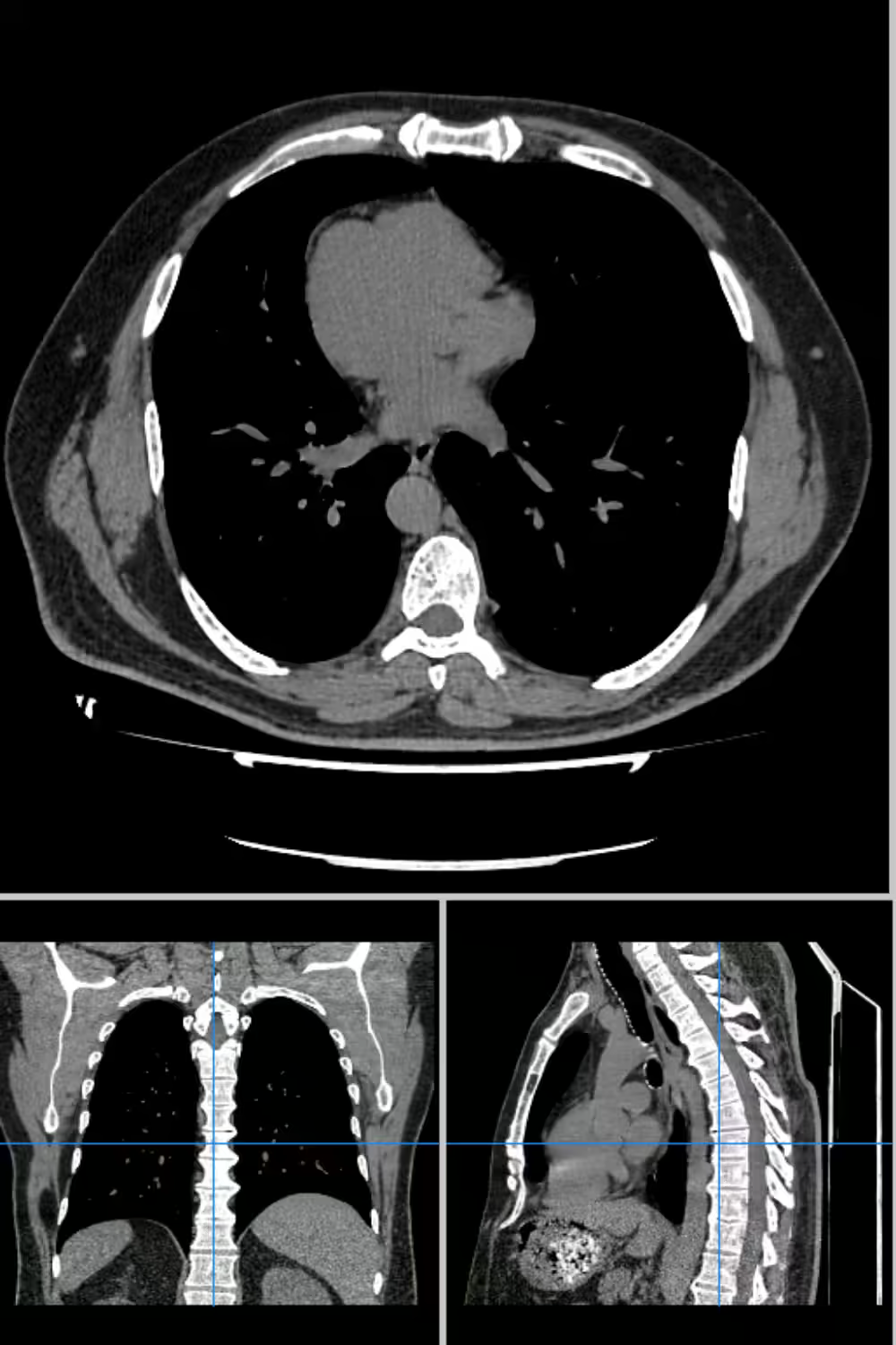
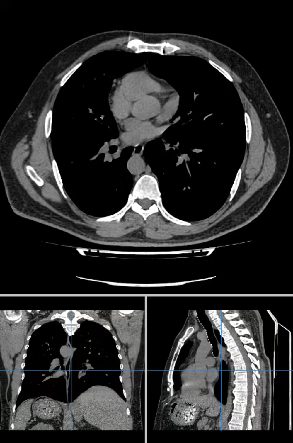
CT Scan Chest Dataset
It is a large-scale CT scan chest dataset featuring over 150,000 chest CT images with annotated pathologies, designed for training deep learning models in lung disease detection, cancer diagnosis, and medical imaging tasks, with labeled data covering a wide range of conditions such as pulmonary embolism, tuberculosis, and lung cancer.
50,000+ studies with protocol
100,000+ studies without protocol
24 pathologies -
 Commercial
- Medicine
- Segmentation
- Computer Vision
- Classification
- Machine Learning
Brain CT Segmentation Dataset
It is a detailed brain CT dataset featuring over 1,000 annotated CT scans for tumor segmentation, brain hemorrhage detection, and other pathology classification tasks, designed to train machine learning models in semantic segmentation, treatment planning, and accurate analysis of brain tissues, structures, and injuries in medical imaging.
1,000+ medical studies
10 pathologies
Why Companies Trust Unidata's Datasets
Share your project requirements, we handle the rest. Every service is tailored, executed, and compliance-ready, so you can focus on strategy and growth, not operations.
What our clients are saying

UniData

Data purchase
Our team got in touch with UniData for purchasing video data. The team at UniData was transparent, timely, and pleasant to communicate and negotiate with. Their samples and descriptions aligned well with the data we received. We will certainly reach out to UniData again if we're in search of 3rd party video data.

Data is well organized and easy to…
Data is well organized and easy to consume. We could download and use it for training within few hours of receiving the data links.
Our Clients Love Us
Ready to get started?
Tell us what you need — we’ll reply within 24h with a free estimate

- Andrew
- Head of Client Success
— I'll guide you through every step, from your first
message to full project delivery
Thank you for your
message
We use cookies to enhance your experience, personalize content, ads, and analyze traffic. By clicking 'Accept All', you agree to our Cookie Policy.