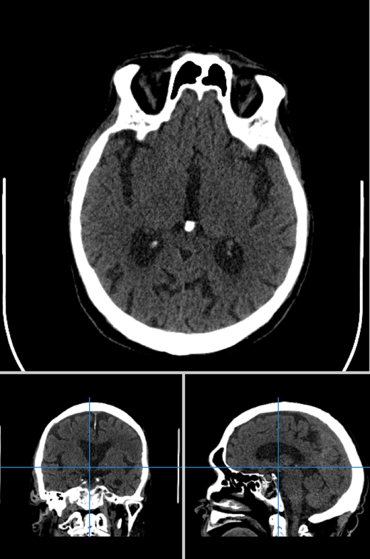
Chest X-Ray Dataset
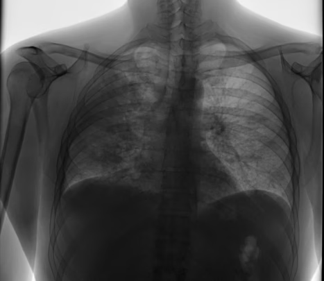
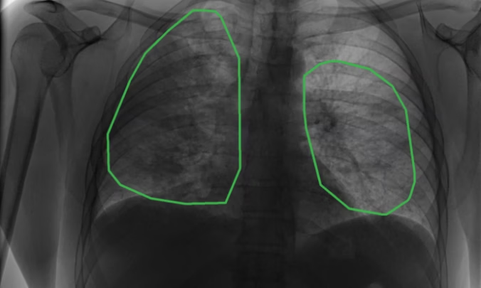
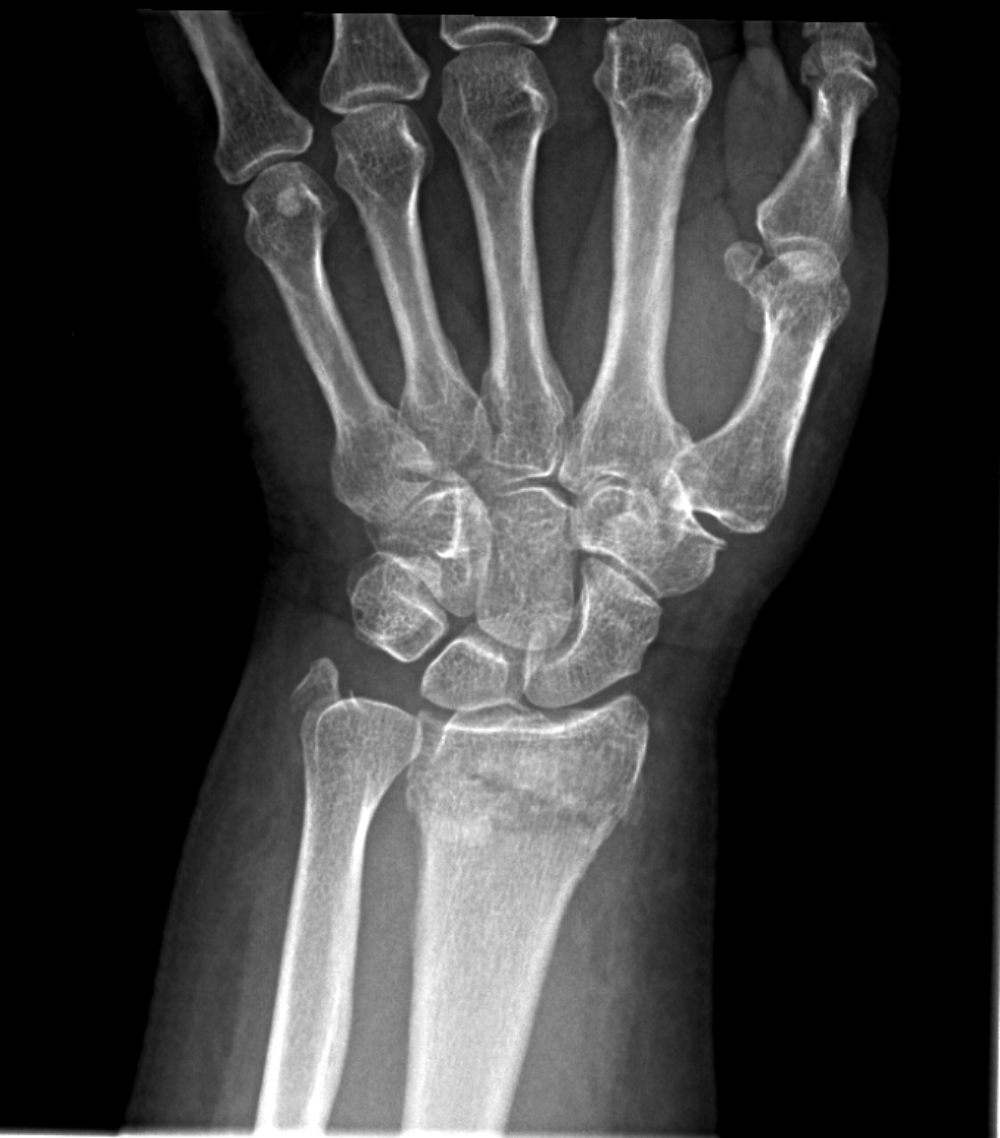
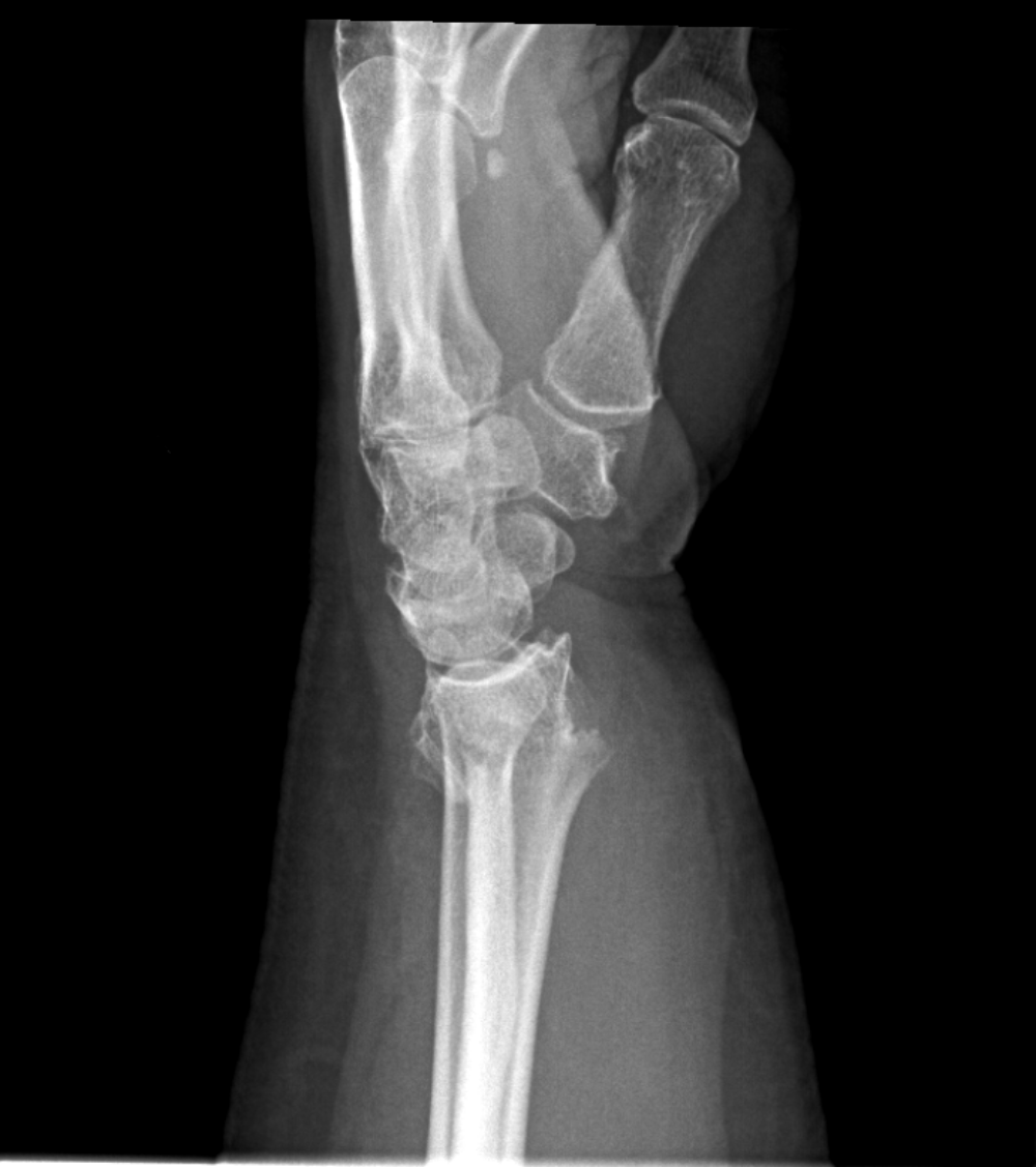
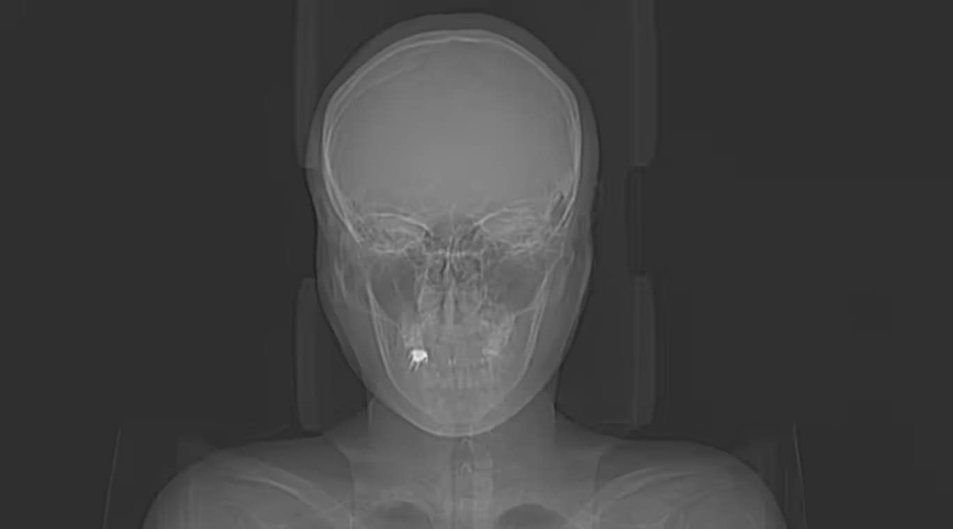
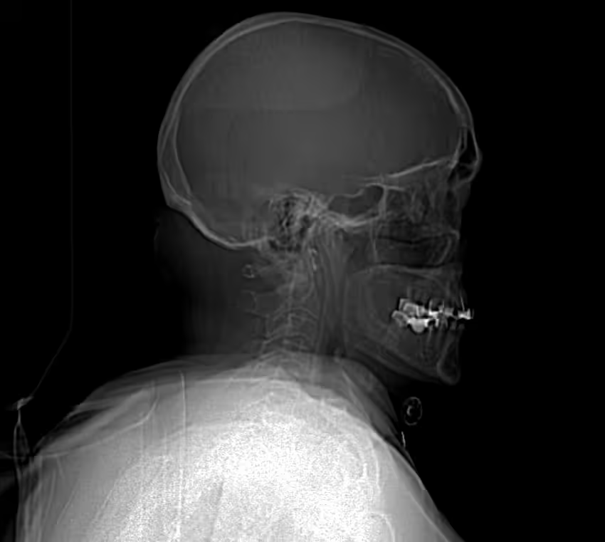
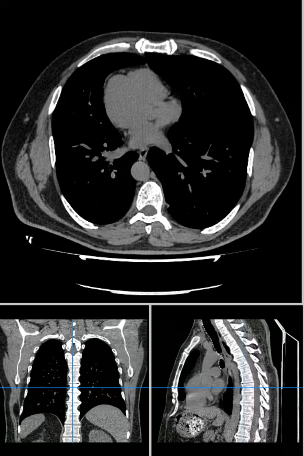
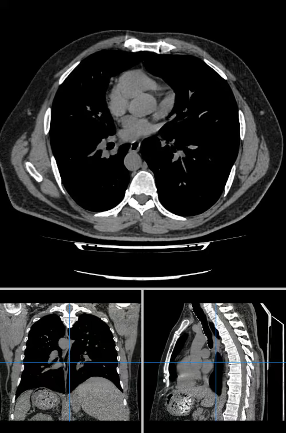
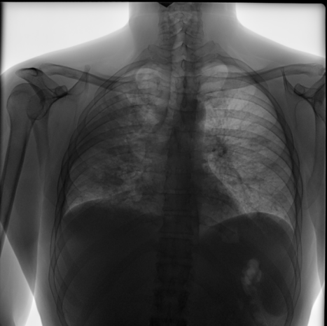
The NIH chest X-ray dataset contains labeled chest radiographs with detailed segmentation of pathologies, providing high-quality X-ray images of chest for detecting lung diseases, pulmonary conditions, and other medical imaging tasks, making it a valuable resource for diagnostic imaging, computer vision, and deep learning in healthcare
-
- Files
- 443
-
- Medical Studies
- 150
-
- Data tags
- 13
- Medicine
- Computer Vision
- Segmentation
- Classification
- Machine Learning
The NIH chest X-ray dataset contains labeled chest radiographs with detailed segmentation of pathologies, providing high-quality X-ray images of chest for detecting lung diseases, pulmonary conditions, and other medical imaging tasks, making it a valuable resource for diagnostic imaging, computer vision, and deep learning in healthcare
- Medicine
- Computer Vision
- Segmentation
- Classification
- Machine Learning
-
- Files
- 443
-
- Medical Studies
- 150
-
- Data tags
- 13
Dataset Info
| Characteristic | Data |
| Description | Chest X-ray to recognize pathologies |
| Data types | DiCOM |
| Markup | Segmentation of pathologies |
| Tasks | Pathology recognition, computer vision. |
| Total number of files | 443 |
| Number of studies | 150 |
| Labeling | ‘Nodule/mass’, ‘Dissemination’, ‘Annular shadows’, ‘Petrifications’, ‘Pleural effusion’, ‘Pneumothorax’, ‘Rib fractures’, ‘Healed rib fracture’, ‘Atelectasis’, ‘Enlarged mediastinum’, ‘Hilar enlargement’, ‘Infiltration/Consolidation’, ‘Fibrosis’ |
| Gender | Male, female |
| Age | 25 - 70 |
Statistics
-
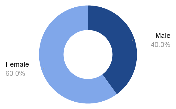
- Distribution by gender
-
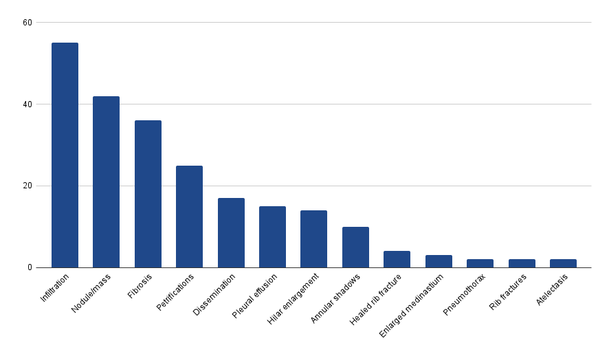
- Number of studies for each condition
Technical
Characteristics
| Characteristic | Data |
| File extension | DiCOM |
| Markup format | JSON |
Dataset Use Cases
FAQs
Unidata Cases
Similar Datasets
Why Companies Trust Unidata's Datasets
Share your project requirements, we handle the rest. Every service is tailored, executed, and compliance-ready, so you can focus on strategy and growth, not operations.
What our clients are saying

UniData


Our Clients Love Us
Ready to get started?
Tell us what you need — we’ll reply within 24h with a free estimate

- Andrew
- Head of Client Success
— I'll guide you through every step, from your first
message to full project delivery
Thank you for your
message
We use cookies to enhance your experience, personalize content, ads, and analyze traffic. By clicking 'Accept All', you agree to our Cookie Policy.
